
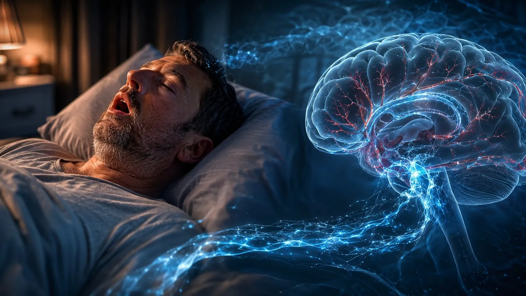
Sleep apnoea brain: why snoring stole 28 nights of brain cleansing.
Sleep apnoea brain — this is not a metaphor. A new study using 7-tesla MRI has shown that sleep apnoea weakens the mechanism by which the brain flushes itself out at night. And it's not sleep fragmentation that's to blame, as previously thought. Falling blood oxygen levels are to blame.
Gawon Cho’s team scanned 36 people: 28 with newly diagnosed obstructive sleep apnoea and 8 without it. Every hour with an oxygen level below 90% was associated with weaker synchronisation between brain pulsations and cerebrospinal fluid flow. It is precisely this synchronisation that removes waste from the tissue. Respiratory micro-awakenings without a drop in oxygen levels did not produce this effect at all.
What is sleep apnoea brain and why is it about waste
The brain does not have lymphatic vessels in the conventional sense. Instead, a glial system operates – a network of spaces around the vessels where cerebrospinal fluid bathes the tissue and removes metabolic waste. Among this waste are beta-amyloid and tau, the same proteins that accumulate in Alzheimer's.
The system is most active during sleep, especially in the deep slow-wave phase. During this time, the spaces between brain cells expand, and fluid can flow more easily. The engine of this flow is arterial pulsation. Each heartbeat creates a pressure wave, fluid moves, and waste is cleared. When pulsation and fluid influx are coordinated out of phase, the cleaning process works: a vessel constricts – fluid enters, compensating for the volume.
Sleep apnoea damages this very coordination. And it does so in a specific way that research has, for the first time, clearly separated into two distinct mechanisms - hypoxia and sleep fragmentation.
Hypoxia, not fragmented sleep
It was previously thought that apnoea harmed the brain primarily through constant micro-arousals – sleep is fragmented, deep sleep phases are reduced, and clearance suffers. This is a logical assumption, but the Cho study revealed something different.
The participants underwent polysomnography or a WatchPAT test. Two parameters were measured separately. T90 is the time spent with oxygen levels below 90% SpO₂, which represents hypoxic stress. RERA — respiratory micro-arousals without a drop in oxygen levels, which is fragmentation. Each participant then underwent a 7T fMRI scan at rest to measure the coupling between grey matter oscillations and fluid influx.
The result is clear. A larger T90 predicted poorer coordination of pulsations and fluid flow (β=0.08, p=0.03), independently of arousals and demographic adjustments. RERAs, however, did not correlate with either pulsation coordination or strength at all. In other words, the sleep apnoea brain suffers from a lack of oxygen, rather than from fragmented sleep. Compare two scenarios. Apnoea with mild desaturation and apnoea with deep oxygen dips represent different burdens on the brain. This is true even if the number of arousals per night is the same.
Why does oxygen weigh more than waking
The mechanism is plausible. Repeated drops in oxygen damage the walls of the brain's blood vessels. A stiffer vessel pulsates less well and draws in less compensatory fluid. Pulsation seems to be present, but it no longer removes waste – it's running on empty.
This is evident in the numbers. In areas where hypoxia increased pulsation amplitude, this amplitude no longer provided a stronger grip on the fluid flow. And where hypoxia did not affect pulsation, stronger pulsation, as expected, led to better clearing (β=−0.004, p<0.001). The difference between these two scenarios is statistically significant.
This is most pronounced in the temporal lobe. It is there that during apnoea the pulsations were elevated but detached from the compensatory movement of fluids. The temporal lobe is one of the first areas to suffer from Alzheimer's.
Why does severe sleep apnea hit the brain harder
The effect is scaled with severity. In individuals with severe apnoea (AHI index ≥ 15 events per hour), the association between hypoxia and impaired coherence was even more pronounced. This remained the case after adjusting for awakenings (β=0.0007, p=0.001). This group statistically differed from individuals without severe apnoea (Z=2.42, p=0.02). It also differed from brain regions where hypoxia did not increase pulsation.
This means that sleep apnoea brain is not an on/off switch, but a gradient. The longer and deeper the oxygen drops at night, the more the nightly cleaning becomes desynchronised. Mild apnoea is also not safe, but severe apnoea works on the brain more methodically.
How is this related to dementia?
Cho's research gives meaning to long-standing epidemiological data. People with untreated apnoea have a higher brain amyloid burden and a significantly increased risk of dementia. A mechanism had long been missing: why apnoea specifically, and not just «poor sleep».
Now the mechanism is specific. This is no longer a general «lack of sleep is harmful», but a concrete chain. Hypoxia toughens brain vessels, stiff vessels pulsate worse and draw fluid poorly. Night-time amyloid clearance weakens, protein builds up over years. A separate line of research shows something important. Even one night of sleep deprivation already changes the physiological pulsations of the brain. The system is sensitive to the sleep state quickly, and sleep apnea brain puts pressure on it every night.
This is not a diagnosis or a sentence. But it is an argument to take your own snoring seriously, especially after 50, when the risk of neurodegeneration is already increasing.
What does this mean in practice
Firstly: sleep apnoea needs to be diagnosed, not simply put up with. Snoring with pauses, morning headaches, daytime sleepiness, a foggy head, and trouble finding the right words in the morning. This isn’t a personality trait or a sign of ageing. It’s a sign that you need to get checked out. Diagnosis is carried out using polysomnography in a sleep laboratory or a home test such as WatchPAT. The gold standard of treatment is CPAP, a continuous positive airway pressure device that keeps the airways open and prevents oxygen desaturation. The problem is well known: up to 30–80% of people stop using CPAP. But it is precisely the hypoxia that it eliminates that is the main driver behind these figures.
Secondly: sleep duration alone isn't the answer here. You can sleep for eight hours and still not get enough cleansing if your oxygen levels drop hundreds of times a night. The amount of sleep and its quality in terms of oxygen levels are different things. A tracker showing «8 hours of sleep» knows nothing about your desaturation.
Third: movement and vessels. The health of the brain arteries is what training directly affects. Aerobic activity maintains the elasticity of blood vessels, and it is vascular stiffness that breaks the mechanism in apnoea. Training will not replace CPAP, but it works with the same bottleneck – the vascular wall. More on how movement flushes the brain can be found in our analysis of the glial system. And we wrote about how physical activity rewrites the brain after 50 Here.
What research has not yet proven
Honest disclaimer. This is a pre-print that has not been peer-reviewed. The sample size was small: 36 people, of whom only 8 did not have apnoea. The design is cross-sectional: it shows a link at a single point in time, not causation. From these data, we do not know if CPAP restores pulse coordination, as treated and untreated groups were not compared separately. Also, 7T MRI is a research tool for now, not something a local GP would prescribe.
But the direction aligns with the broader picture. Falling oxygen levels stiffen blood vessels, stiff vessels pump fluid less efficiently, and nightly cleansing relies precisely on this flow. The power of this work isn't in sample size. It's in the fact that the study separated two mechanisms that were previously conflated. And it showed the main point: hypoxia is to blame, not arousal.
Sleep apnea brain — a working hypothesis with a specific, measurable mechanism. And another reason not to ignore your own snoring, but to get checked before waste builds up over the years.
Sources
- Cho G, Kam K, Chen A, et al. Greater hypoxic burden predicts weaker coordination between brain pulsation and CSF flow on 7T MRI independent of non-hypoxic arousals: Implications for glymphatic activity. bioRxiv. 2026. DOI: 10.64898/2026.04.25.720853
- Helakari H, Järvelä M, Väyrynen T, et al. Effect of sleep deprivation and NREM sleep stage on physiological brain pulsations. Frontiers in Neuroscience. 2023;17:1275184. DOI: 10.3389/fnins.2023.1275184
- Wang X, et al. Impact of Sleep Disorders and Disturbed Sleep on Brain Health: A Scientific Statement From the American Heart Association. Stroke. 2024. DOI: 10.1161/STR.0000000000000453






 Українська
  English (UK)